KING EDWARD MEMORIAL HOSPITAL
SETH GORDHANDAS SUNDERDAS MEDICAL COLLEGE
बृहन्मुंबई महानगरपालिका रुग्णालय
KING EDWARD MEMORIAL HOSPITAL
SETH GORDHANDAS SUNDERDAS MEDICAL COLLEGE
बृहन्मुंबई महानगरपालिका रुग्णालय
The Department of Dermatology was established at Seth GS Medical college and KEM Hospital asold as the hospital by Dr. A C Rebello in 1926 at Ward No.17&18 Main Hospital Bldg & Gr. Floor.
From 1926 Seth GS Medical college had post of honorary dermatologist and venereologist and honorary lecturer, with extern to assist outpatient department. With increased work appointment of resident registrar in 1930.In 1931 house physician was appointed. Honorary dermatologist started lectures and clinical demonstrations .
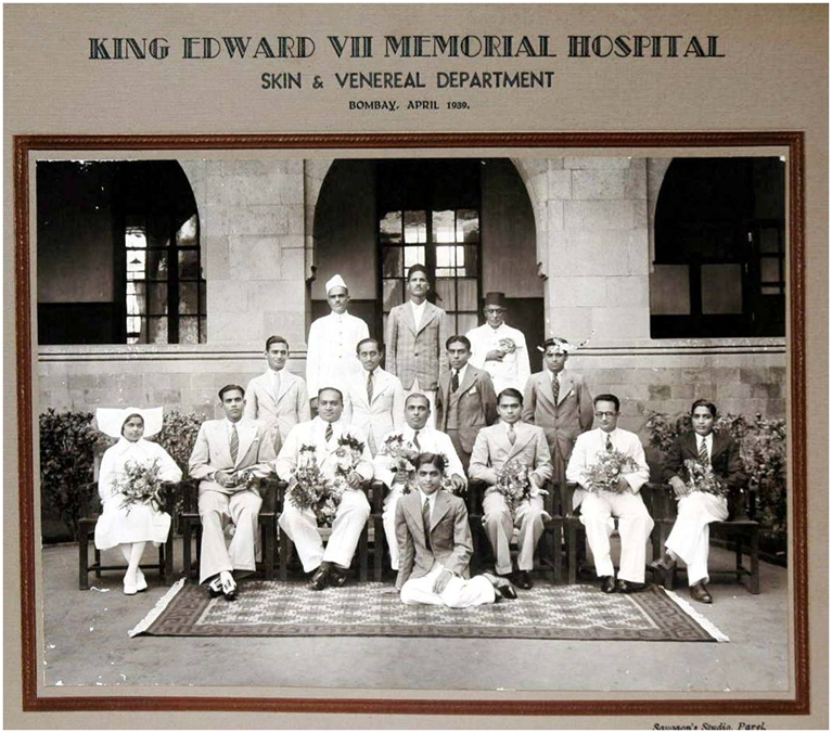
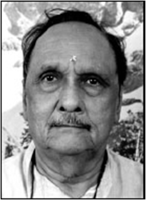
Later Dr. Sharat Desai established first separate Department of dermatology in KEM hospital.
He created first residency program in dermatology.
He contributed in mycology and initiated dermatopathology.
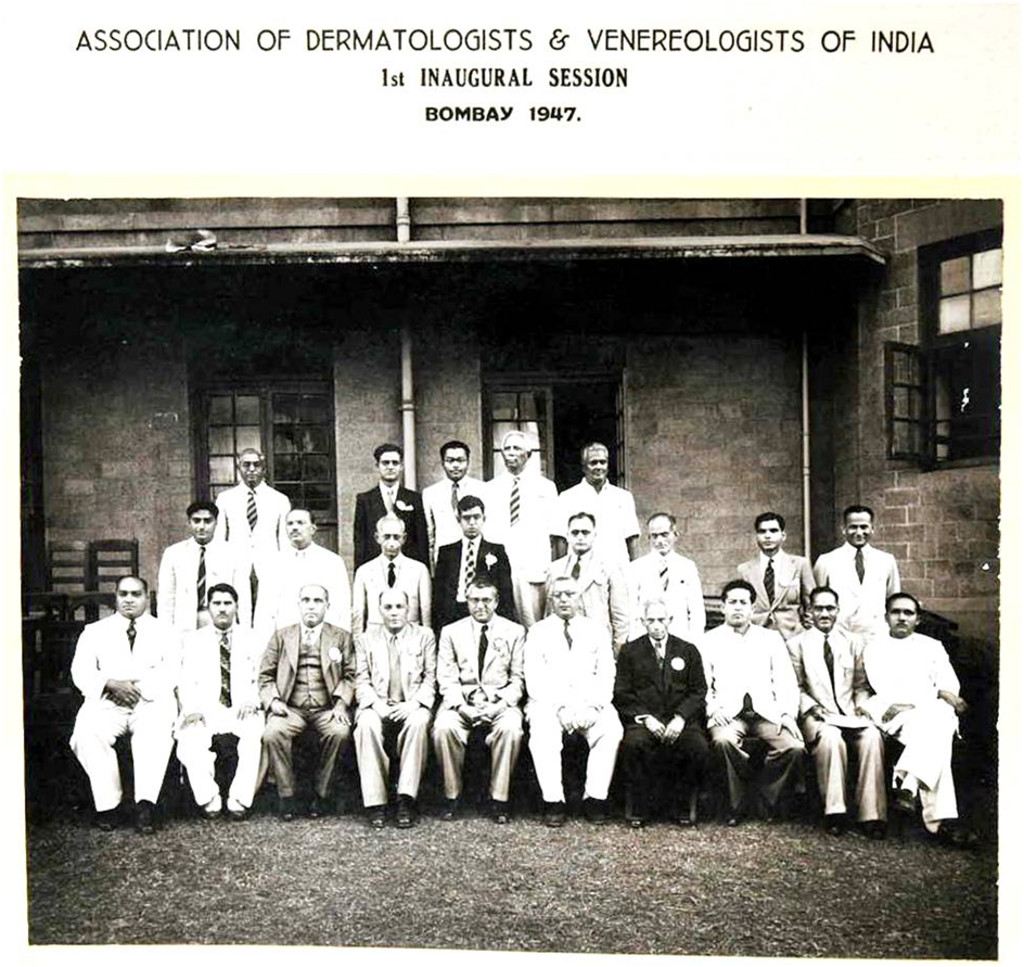
On July 1st 1947 Dr. UB Narayan Rao with Shri Morarji Desai ( home and revenue minister) inaugurated IADVL Mumbai.
Seth GS Medical college hosted this inaugural function.
In 1980s Dr. Rui Fernandez initiated specialty dermatology clinics. Later in 1989 he was Organizing Secretary for National Conference in Mumbai.
In 1990s he also pioneered dermatosurgery and paediatric dermatology speciality.
In 2004 he was President of IADVL.
From 1996 to 2000Dr. Pushpa Gupte was Head of Department of Dermatology.
In 2000 she organized first All India Dermatomycology CME and took keen interest in developing dermatopathology.
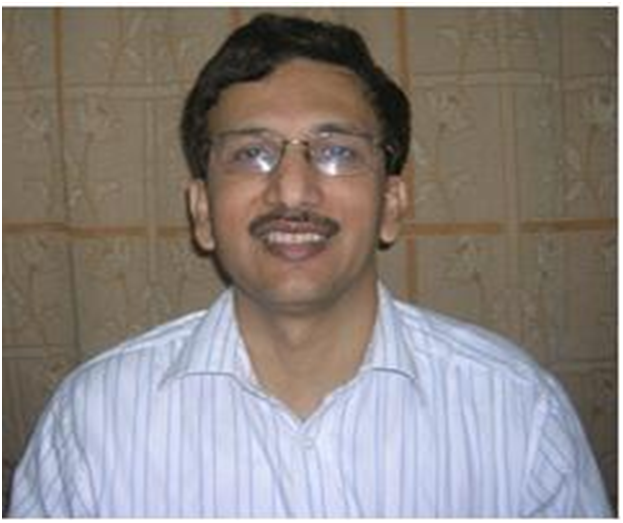
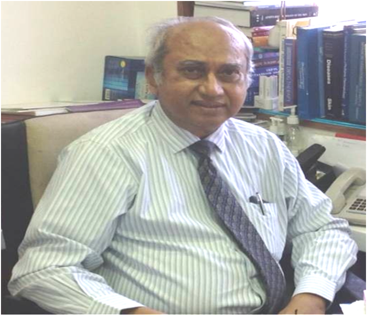
In 2002 Dr. Uday Khopkar joined Department as Professor and Head of Department.
He initiated Ultraviolet therapy , Dermoscopy, Pulse therapy for vesiculobullous disorders and Laser therapy in institute.
From 2003 to 2008 he was chief editor of IJDVL
He is Honorary Dermatologist to the Governor of Maharashtra
He is founder and Guide for Fellowship in Diagnostic Dermatology Course at MUHS in India
He is teacher and examiner for postgraduate students in degree and diploma courses in dermatology.
Scientific Chairperson for the National Conference of IADVL – 2004, Asian dermatology congress 2016
Organized CMEs in Dermatopathology, Dermoscopy, Genodermatosis, Update in Clinical Dermatology, Histopathology of Leprosy, Update in Leprosy
Chief Editor of Indian Journal of Dermatology, Venereology and Leprology for 6 yrs 2003-08
Section Editor of API Textbook of Medicine 2008 (printed the whole section in color for the first time)
Authored books on
Skin and STDs,
HIV infection,
Drugs in dermatology,
Dermoscopy of pigmented skin and
Skin biopsy
Lichen planus
Initiated and popularized IJDVL e-library project 2009
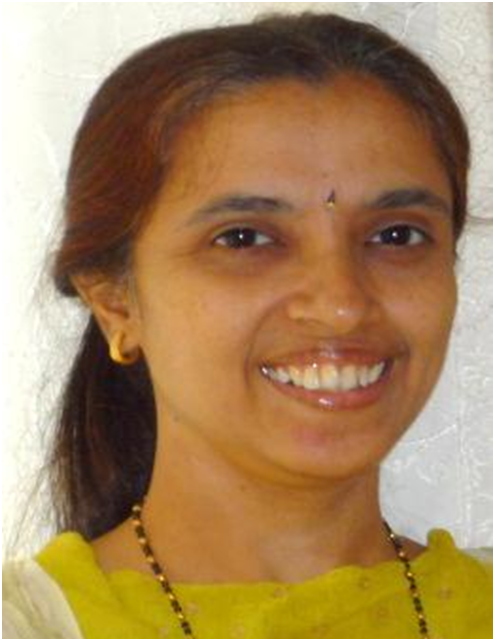
In 1991 Dr. Vidya Kharkar joined Department as lecturer, later became Associate professor and at present she holds the post of Professor. She haskeen interest in clinical dermatology including paediatric and geriatric dermatology. She started Non cultured melanocyte grafting for vitiligoand PRP for alopecias.
She completed fellowship in histopathology from CMC, Vellore and HIV from Mumbai.She is exeutive member of Lupus India Forum Ensemble(LIFE).She is on editorial review board for variousjournals . Co authored chapter in IADVL. text book of dermatology.
In 1993 she joined as lecturer in Department of Dermatology with keen interest in clinical dermatology- Acne,Psoriasis, Vitilligo
She is undergraduate and postgraduate teacher and examiner of diploma and degree courses in dermatology for MUHS and University of Mumbai.
She is exeutive member of Lupus India Forum Ensemble(LIFE)
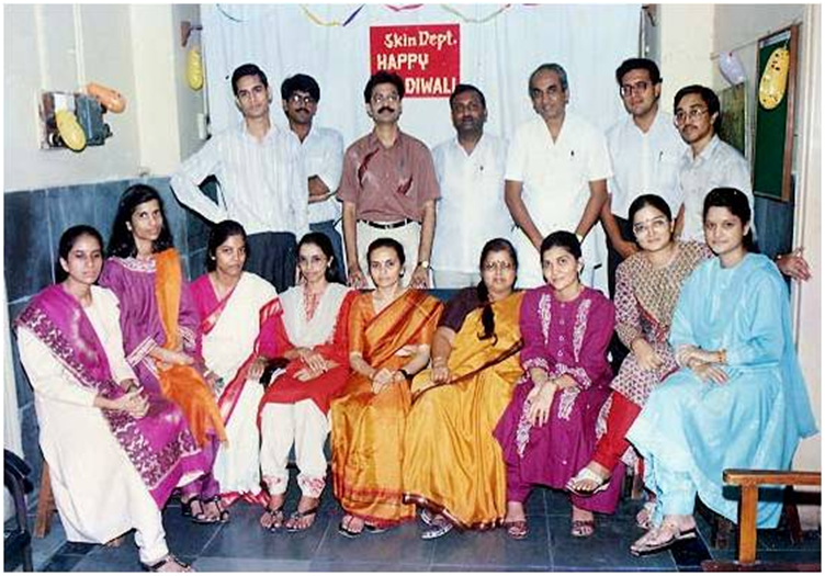
CME on Leprosy on occasion of 90 yrs of completion of Seth GS Medical College and KEM Hospital
Dr. Vidya D. Kharkar, M.D. (Skin), D.V.D.
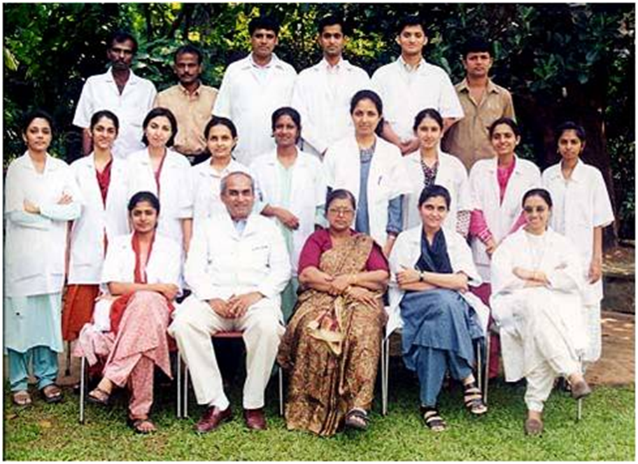
Department Staff :
| Dr. Vidya D.Kharkar | Prof & Head |
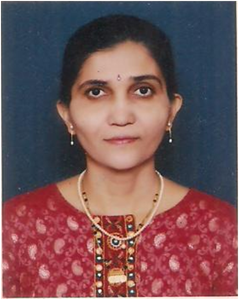
| Dr. Sunanda A Mahajan | Additional Professor |
| Dr. Siddhi Chikhalkar | Associate Professor |
| Dr. Prachi Gole | Assistant Professor |
Present Position of Residents :
| Fellows | 2 |
| BSR | 2 |
| Senior Residents | 1 |
| Junior Residents | 16 |
| Clerk cum Typist | 1 |
| Lab. Technician | 1 |
| Staff Nurse-OPD | 1 |
Group Lectures for all 2nd & 3rd MBBS students.-CVTC Auditorium.
For undergraduate students :
Posted for skin term in the department.
For OT/PT students :
Lecture series yearly
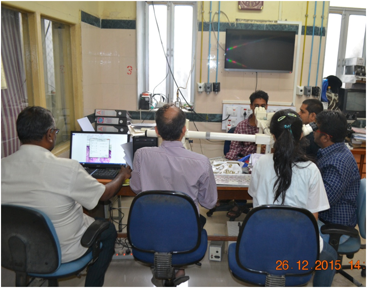
For Post graduate students
Dermato Pathology :
Clinicopathological correlation and discussion of cases – Every Tuesday & Saturday
Grand Round– Monday
Seminars –Wednesday 2-3pm
Histopathology Seminars- Tuesday 2-3pm
Journal Club –Thursday 2-3pm.
Case Presentation– Fri 2-3 pm
P.G. Registration :
Routine Daily OPDs :
Special clinics
Cosmetology clinic – Every Monday
Vitiligo clinic – Every Tuesday
Psoriasis clinic – Every Wednesday
Paediatric dermatology clinic – Every Thursday
Acne clinic – Every Friday
STI Clinic – Daily
Leprosy clinic- Tuesday and saturday
Vesicobullous diseases speciality clinic – every tuesday and thursday
Hair clinic- Saturday
Autoimmune diseases speciality clinic- Daily
School clinic:- 1pm to 3pm
Dematosurgical procedures:-On Tues/Sat
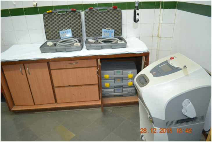
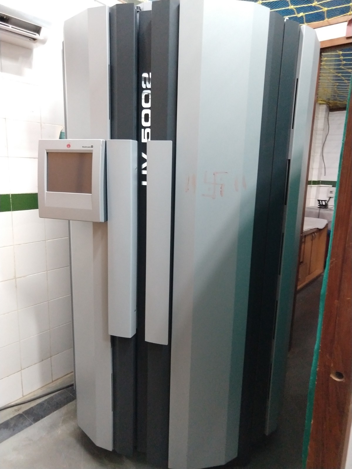
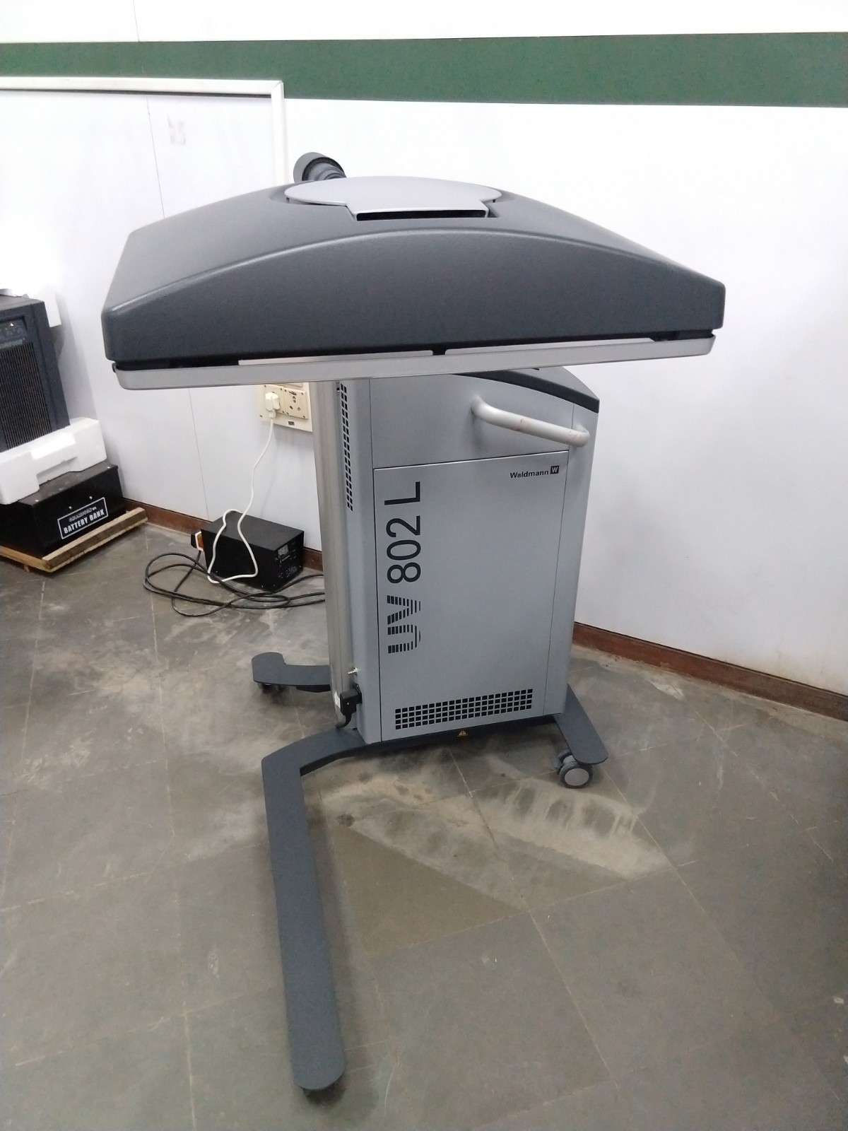
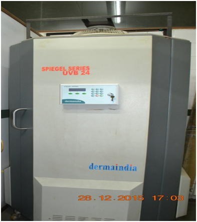
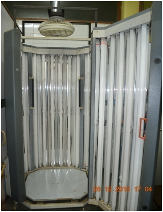
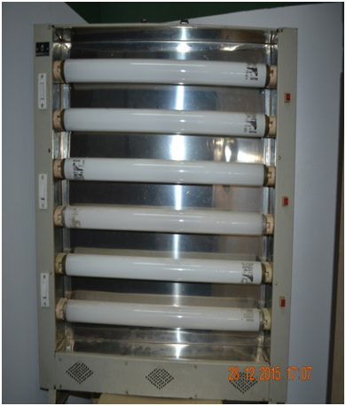
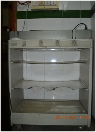
Phototherapy
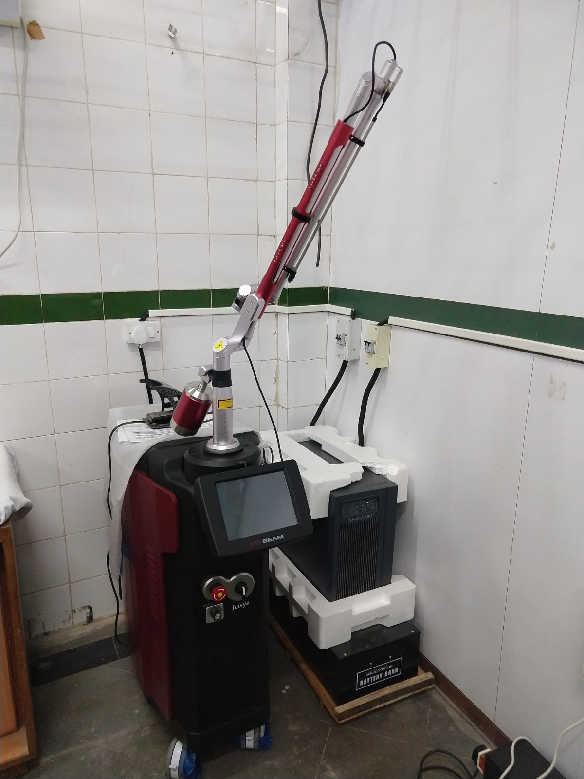
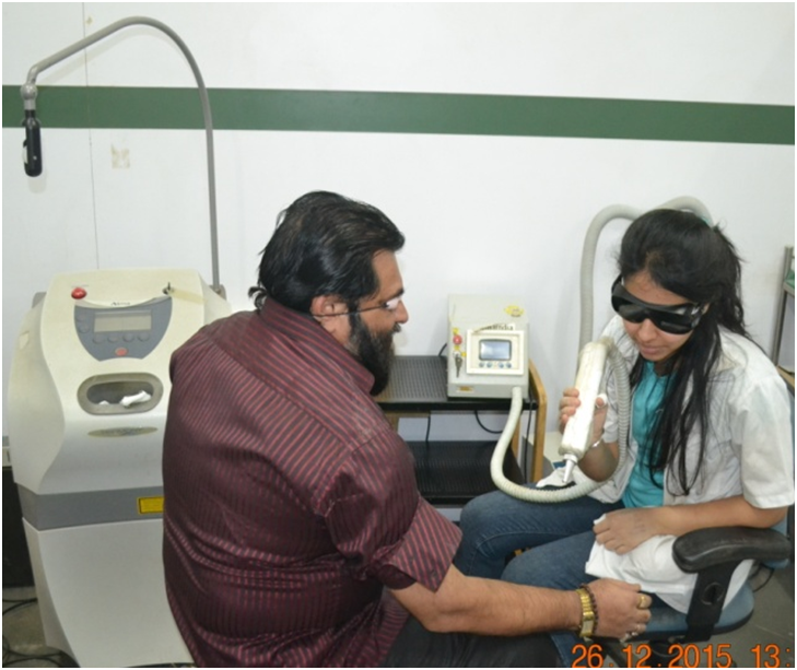
Lasers
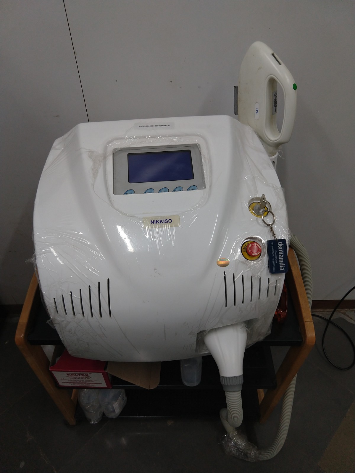
The department is equipped with multiple lasers used for the treatment of various skin conditions at minimal cost.
Tribeam Q switched laser- Monday/ Wednesday/Saturday 1-3 pm
Intense pulse light (IPL)- Tuesday/ Thursday 1 to 2 pm.
Excimer laser (308nm)- every tuesday after vitiligo clinic(12.30pm)
Dermatopathology services
Patients with doubtful diagnosis are biopsied for confirmation of diagnosis. The department has a state of the art multiheaded microscope with fluorescence attachment. The microscope is equipped with teaching heads and a TV monitor for teaching postgraduates. It is also connected to a PC for storing still images. The department receives a lot of references of difficult cases for dermatopathology opinion and clinicopathological correlation. The department also has a laboratory where biopsy samples are processed.
Dermatoscopy
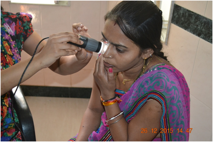
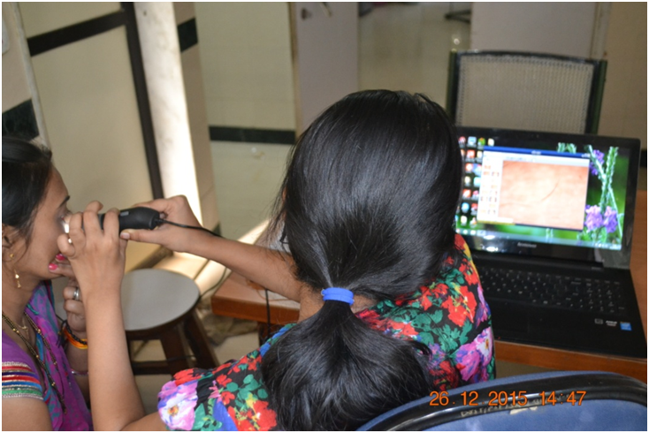
Department has a Dermatoscope which is connected to a computer. It provides for non invasive evaluation of various skin conditions. Images are stored on the computer for correlation with clinical and histological findings and can be retrieved at a later date for comparison
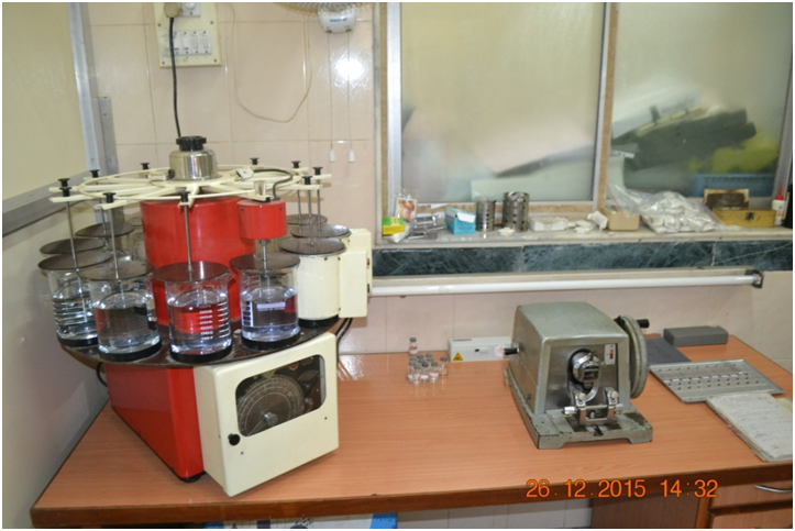
Mycology services
| Q switched NDYAG | IPL |
| TATTOO REMOVAL: | HAIR REMOVAL |
| LENTIGENES/ FRECKLES | KELOID |
| NEVUS of OTA & ITO: | ACTIVE ACNE |
| MELASMA / LPP | VASCULAR LESIONS |
| LASER TONING/ CARBON PEEL |
| COSMETOLOGY CLINIC | Monday |
| VITILIGO CLINIC | Tuesday |
| PSORIASIS CLINIC | Wednesday |
| PEADIATRIC DERMATOLOGY: | Thursday |
| ACNE CLINIC | Friday |
| HAIR: | Saturday |
| HENSENS CLINIC | Tuesday,Friday |
(Chemical cautery, Needling, ILS, Comedone extraction, Molluscum extraction, Parring, Podophyllin application, TCA application,wound dressing)
VITILIGO: Suction blister
Punch grafting
Dermabrasion
Shave graftig
HAIR: Follicular unit transplantation
LIPOMA EXCISION
SEBACEOUS CYST EXCISION
NAIL AVULSION
ACNE SCAR REVISION
PHENOL APPLICATION (Alopecia,Vitiligo)
DPCP (Alopecia Areata)
CRYOTHERPY
ELECTROCAUTERY
RADIOFREQUENCY ABLATION
PRP WITH DERMAROLLER
PATCH TEST (to rule out allergies):
ASST/AST (for chronic idiopathic urticaria)
BIOPSIES
WOODS LAMP FOR DIAGNOSIS
Peels for Acne vulgaris &postinflammatory hyperpigmentation
For facial rejuvenation & melasma
Blood Collection for VDRL & HIV Tests
KOH Mount & Culture for Fungus
Hair mount
Staining of smears for microorganisms
Smears for bullous skin diseases
Dermatopathology Reporting
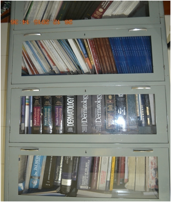
SEMINAR ROOM WITH LIBRARY
A 28-year male presented to the skin clinic for progressive cutaneous lesions of the neck that he had for the past 3 years. He was diagnosed as a case of Wilson’s disease 15 years back and started on D penicillamine at a dose of ~800mg daily. He was asymptomatic for 13 years when he developed mild itchy lesions starting over nape of neck which progressed to involve bilateral axilla gradually. Clinical examination revealed multiple match head sized keratotic papules on an annular erythematous base on nape of neck and axilla. Also we noticed skin over axilla was pendulous and hanging in folds. The clinical and histopathological findings were collaboratory.
A 56 year/Male patient of UP presented with multiple asymptomatic,hypopigmented maculesof varying sizes all over body since 6 months and multiple firm to hard painless nodules ranging from 1-5 cm on extremities since 3 months. Biopsy from the macule showed a sparse superficial perivascular infiltrate of lymphocytes and histiocytes and the nodule showed diffuse dense infiltrate involving the upper and mid dermis with tiny slightly basophilic intracytoplasmic dots of size 1-2 microns.
Visit on 29 May 23 by Professor Joseph Muenzer, Pediatric Geneticist , University of North Carolina
Workshop on “Principles of Scientific Writing” 12, 13 October 2017, conducted for Pfizer Ltd
Mr Ashnik Chauhan at the DIA meeting in Chicago in June 2017
90 years of Seth GS Medical College and KEM Hospital, January 2016